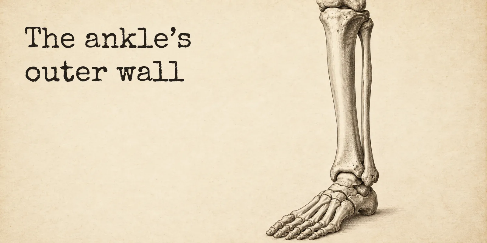
The fibula takes a small share of the load through your lower leg, about 6 to 17 percent in cadaver studies, and the textbooks round that to none. Its lower end is the outer wall of your ankle joint, and that position matters more than the weight.
The fibula does bear some weight, though not much. Studies that loaded legs from donated bodies found it taking roughly 6 to 17 percent of the force through the lower leg, with the tibia beside it carrying the rest. Your ankle needs the fibula for a different reason, which is that its lower end forms the outer wall of the ankle joint.
I came to this from my own lab. The field note The Bone Room gives for the fibula says it carries “little or no body weight,” and when I went back to the references, OpenStax says the fibula does not bear weight and StatPearls calls it not a weight-bearing bone. So the lab said little, the textbooks said none, and I wanted to know whether the little was real.
Two bones in the leg, and one does the carrying
Anatomy keeps the word leg for the stretch between the knee and the ankle, and there are two bones in it. The tibia, your shinbone, sits on the inner side and is the main weight-bearing bone of the lower leg, as well as the second longest bone in the body. The fibula runs down the outer side, and OpenStax’s word for it is slender.
The name suits it. Fibula was Latin for a clasp or brooch, and English has used it for the smaller bone of the lower leg since 1706, on the idea that the pair resembles a clasp, the same kind of fastening a safety pin still uses. You can see the two of them together, with the femur and the kneecap above, on the bones of the leg.
The textbooks say none, and the measurements say some
When researchers want a number, they fit force gauges into legs from donated bodies and press down. Three of those studies give figures you can compare.
| Study | Ankle position | The fibula’s share of the load |
|---|---|---|
| Takebe and colleagues, 1984 | Neutral | 6.4% |
| Goh and colleagues, 1992 | Neutral | 7.12% |
| Wang and colleagues, Oxford, 1996 | Axial load of 1,500 newtons, ten limbs | 17% on average |
A review in the Journal of Athletic Training goes wider still and puts the fibula at 10 to 30 percent of a static load. Some of the spread comes from the ankle itself, because all three lab studies found the fibula’s share going up in dorsiflexion, the position where the top of your foot comes up toward your shin. Takebe’s and Wang’s teams also found it going down when the foot pointed away.
I don’t think the textbooks and the studies are really arguing. OpenStax and StatPearls are saying which bone is built to carry you, and nobody disputes that it’s the tibia. The studies are measuring what passes through the fibula anyway, and none of them came back with zero.
The bump on the outside of your ankle
Press on the outside of your ankle and the hard knob under your fingers is the bottom of the fibula. It’s called the lateral malleolus, and its partner on the inside, the medial malleolus, is the bottom of the tibia.
Between those two knobs sits the talus, the top bone of the foot. The underside of the tibia makes a roof over it and the two malleoli make the walls, and together they’re called the ankle mortise, borrowed from carpentry, where a mortise is the hollow cut into wood to take a matching projection.
OpenStax says the malleoli hold the sides of the talus firmly and prevent any side-to-side motion, and that’s what makes the ankle a hinge that only tips your foot up and down. Take the lateral malleolus away and, according to StatPearls, the mortise loses its stability.
The outer wall is also an anchor. Three ligaments run from the lateral malleolus, the anterior and posterior talofibular ligaments across to the talus and the calcaneofibular ligament down to the heel bone. The front one is the weakest of the three, and it’s the ligament most often injured when an ankle is sprained.
How the two bones are tied together
For most of their length, the tibia and fibula are joined by the interosseous membrane, a sheet of fibrous tissue stretched between a narrow ridge on each bone.
Near the ankle the connection tightens into a syndesmosis, a word that means fastened with a band. OpenStax describes it as strongly uniting the two bones and allowing little movement, and it’s what locks the talus in place between them. Ligaments at the front, at the back and between the bones do most of the fastening.
The membrane also shapes where the fibula’s share of the load ends up. Wang’s team found it changing how load was split between the two bones, with the distal fibula, the end farther from the hip, carrying a higher proportion than the upper end.
What the fibula does when you stand and step
The top of the talus is wider at the front than at the back, by 4.2 millimeters on average. When you bring your foot up toward your shin, that wider front part wedges between the two malleoli, which StatPearls says makes dorsiflexion the most stable position your ankle has.
For the wedge to fit, the fibula has to make room. As the ankle dorsiflexes it rotates outward by around 3 to 5 degrees, and the ligament between the two bones works like a spring, in one review’s description, letting them separate slightly.
It moves down, too. With weight on the leg, the fibula has been measured sliding an average of 2.4 millimeters toward the foot, which deepens the mortise and pulls the membrane taut. Wang’s loaded limbs showed the same direction of travel, the lateral malleolus dropping relative to the medial one.
I’d been picturing the fibula as a strut bolted to the side of the tibia, so the turning and the sliding were the part that surprised me most.
Why one millimeter at the ankle counts
The fit between the talus and the mortise is close enough that very small shifts matter. In a 1976 study of 23 dissected ankles, moving the talus one millimeter sideways cut the contact area between it and the tibia by an average of 42 percent, and most of the loss came in that first millimeter.
Less contact means the same load pressing into a smaller patch of joint. The fibula’s outer wall is part of what keeps the talus centered, and a review of syndesmosis injuries calls restoring the fibula’s length, its alignment and its spacing from the tibia critical to a good outcome.
Surgeons take the middle and leave the ankle end
Surgery gives the clearest picture of how much the leg can spare. Part of the fibula can be used to rebuild a damaged jaw, and the lab’s field note on that says it’s possible because the tibia handles the main weight-bearing job.
When surgeons take bone from the fibula, they take the middle. StatPearls describes the central portion being harvested, with about 5 centimeters left at the bottom for ankle stability and 5 at the top to protect a nerve that crosses the outside of the fibula’s neck. A 2025 systematic review gives the guidance as 6 to 8 centimeters left at the ankle end instead. The two figures differ, and both of them keep the lateral malleolus where it was.
That review, which looked at people who had the operation for head and neck cancer, found ankle instability reported in 3.3 percent of patients and gait disturbance in 5.5 percent. It also points out that the studies it pooled measured these things in very different ways, so the figures are a rough guide.
Where on the fibula a break sits
On a report, “fibula fracture” on its own tells you surprisingly little. Where along the bone the break is, and what else was hurt with it, changes what it means.
A break through the shaft, away from the ankle, is usually the simpler case. StatPearls says the shaft isn’t a weight-bearing portion of the bone, that isolated shaft fractures are rare and mostly come from a direct blow, and that the vast majority are treated without surgery, often with patients allowed to put weight on the leg as they can tolerate it.
A break at the lateral malleolus is filed differently. It’s part of the fibula, but it counts as an ankle fracture and follows different treatment principles. StatPearls also warns that a fibula fracture within 15 centimeters of the ankle joint is likely to come with damage to the tibia, the ligaments or both.
For breaks near the ankle, StatPearls sorts them by where the fibula gives way relative to the syndesmosis, in what’s called the Weber classification.
| Weber type | Where the fibula breaks | What StatPearls says about it |
|---|---|---|
| A | Below the syndesmosis | Typically stable, and can often be treated conservatively |
| B | At the level of the syndesmosis | Can be treated conservatively if stable; unstable ones need surgical fixation |
| C | Above the syndesmosis | Usually unstable, and requires surgical fixation |
One break sits right up near the knee and is still an ankle injury. A fracture at the neck of the fibula, with no other break in the fibula or the tibia, is treated as a telltale sign that the syndesmosis down at the ankle has been torn, and it has its own name, the Maisonneuve fracture.
None of this is a guide to what you should do with your own leg. Whoever is treating you is looking at the whole injury, the alignment of the ankle and the state of the ligaments included, and a label on a report can’t carry all of that. If you’re further along, how a broken bone heals covers what happens next.
Non-weight-bearing means two different things
The phrase turns up in two places. In a textbook, saying the fibula doesn’t bear weight is a description of the bone. On your paperwork after an injury, non-weight-bearing is an instruction about your whole leg.
StatPearls defines the instruction: non-weight-bearing means the leg should not touch the floor or support any body weight. Above it come toe-touch weight-bearing, where the toes touch the floor for balance only, then partial weight-bearing, then weight-bearing as tolerated, which StatPearls puts at 50 to 100 percent of body weight, and finally full weight-bearing. After surgery, it’s the surgeon who decides which one applies.
Frequently asked questions
Does the fibula bear weight?
A little. Cadaver studies have measured it carrying about 6 to 17 percent of the load through the lower leg, more when the foot is pulled up toward the shin, and one review gives a wider range of 10 to 30 percent. OpenStax and StatPearls both call it not weight-bearing, and both name the tibia as the bone that does the carrying.
Why does the ankle need the fibula?
The bottom of the fibula, the lateral malleolus, is the outer wall of the ankle joint, holding the talus in place from the side and anchoring three of the ankle’s ligaments. StatPearls says the mortise loses its stability without it.
Can you walk on a broken fibula?
It depends on where it’s broken and what else is injured, and your clinician decides. StatPearls says most isolated breaks of the shaft are treated without surgery and patients are often allowed weight as tolerated, while breaks near the ankle count as ankle fractures and follow different rules.
Can you live without your fibula?
Surgeons can remove the middle of the fibula to rebuild other bones, such as the jaw, and leave the ends in place. StatPearls says about 5 centimeters are kept at the ankle end for stability, and a 2025 review gives 6 to 8.
What is the lateral malleolus?
It’s the lower end of the fibula, the hard bump you can feel on the outside of your ankle. Its partner on the inside, the medial malleolus, is the lower end of the tibia, and the talus sits between the two.
The fibula is one of four bones in the lab’s leg case, with the femur, the kneecap and the tibia, and a single line in its field note is where this post started. Those notes are the part of the lab I look forward to most, and the story of how it began explains why. If you’d like the fibula as a name you know rather than one you look up, the lab won’t let you claim it until you’ve named it on three separate days, the last time typed from memory. It’s free, and you can start in the lab without an account.
Sources
Every anatomical claim above is checked against OpenStax Anatomy and Physiology 2e or StatPearls on the NCBI Bookshelf, the same references the lab uses. The load measurements, the fibula’s movement and the one-millimeter contact figure come from peer-reviewed studies and reviews, named below, and where they give different figures the text says so.
Show all 18 sources
- OpenStax, Anatomy and Physiology 2e, 8.4 Bones of the Lower Limb: the leg as the part between knee and ankle; the tibia as the main weight-bearing bone of the lower leg and second longest bone; the fibula as slender, on the lateral side, and not bearing weight; the interosseous borders; the lateral and medial malleoli; the talus articulating with the tibia and fibula.
- OpenStax, 9.2 Fibrous Joints: a syndesmosis as “fastened with a band”; the interosseous membrane joining the tibia and fibula; the tibiofibular syndesmosis uniting the bones, allowing little movement and locking the talus in place.
- OpenStax, 9.6 Anatomy of Selected Synovial Joints: the malleoli preventing side-to-side motion of the talus; the ankle as a hinge allowing dorsiflexion and plantar flexion; the anterior and posterior talofibular and calcaneofibular ligaments; the anterior talofibular ligament as the most commonly injured.
- StatPearls, Anatomy, Bony Pelvis and Lower Limb, Fibula: the fibula as not a weight-bearing bone; the lateral malleolus articulating with the talus; the absence of a lateral malleolus destabilizing the mortise; the interosseous membrane attaching at the interosseous border.
- StatPearls, Anatomy, Bony Pelvis and Lower Limb: Ankle Joint: the tibia and both malleoli forming the ankle mortise; the talus wider in front, making dorsiflexion the most stable position; the attachments of the three lateral ligaments; the anterior talofibular ligament as the weakest of them.
- StatPearls, Fibula Fractures: the shaft as not a weight-bearing portion; isolated fractures as rare and usually from direct injury; most shaft fractures treated nonoperatively with weight-bearing as tolerated; lateral malleolus fractures categorized as ankle fractures; a fracture within 15 cm of the ankle likely involving the tibia or ligaments; the Maisonneuve fracture.
- StatPearls, Ankle Fracture: the Weber A, B and C types and their usual stability and treatment.
- StatPearls, Fibula Free Flaps: the central portion harvested, with the distal 5 cm kept for ankle stability and the proximal 5 cm to protect the common peroneal nerve at the fibular neck.
- StatPearls, Weight Bearing: the definitions of non-weight-bearing, touch-down, partial, as tolerated (50% to 100% of body weight) and full weight-bearing; the surgeon defining the amount after surgery.
- Takebe K, Nakagawa A, Minami H, Kanazawa H, Hirohata K. Role of the fibula in weight-bearing. Clinical Orthopaedics and Related Research, 1984: 6.4% of the load to the fibula in a neutral ankle, increasing with dorsiflexion and decreasing with plantar flexion.
- Goh JC and colleagues. Biomechanical study on the load-bearing characteristics of the fibula and the effects of fibular resection. Clinical Orthopaedics and Related Research, 1992: 7.12% in a neutral ankle, ten specimens, maximum loads at full dorsiflexion and eversion.
- Wang Q, Whittle M, Cunningham J, Kenwright J. Fibula and its ligaments in load transmission and ankle joint stability. Clinical Orthopaedics and Related Research, 1996: an average of 17% of a 1,500 N axial load in ten limbs; more in dorsiflexion, less in plantar flexion; the lateral malleolus migrating distally under load; the interosseous membrane modifying load distribution, with the distal fibula carrying a higher proportion.
- Norkus SA, Floyd RT. The anatomy and mechanisms of syndesmotic ankle sprains. Journal of Athletic Training, 2001: the fibula carrying 10% to 30% of a static axial load; the talus 4.2 mm wider in front on average; 3° to 5° of lateral fibular rotation with dorsiflexion; 2.4 mm of inferior fibular migration in weight bearing, deepening the mortise and tightening the interosseous membrane; the three syndesmotic ligaments.
- Porter DA, Jaggers RR, Barnes AF, Rund AM. Optimal management of ankle syndesmosis injuries. Open Access Journal of Sports Medicine, 2014: the interosseous ligament acting as a spring to allow slight separation in dorsiflexion; restoring fibular length, alignment and the tibia to fibula interval as critical.
- Ramsey PL, Hamilton W. Changes in tibiotalar area of contact caused by lateral talar shift. Journal of Bone and Joint Surgery, 1976: 23 dissected ankles; an average 42% reduction in contact area with the first millimeter of lateral talar shift.
- Holm S and colleagues. Ankle instability and gait disturbance after free fibula flap reconstruction in head and neck cancer reconstruction: a systematic review. JPRAS Open, 2025: 6 to 8 cm of distal fibula kept for ankle stability; ankle instability 3.3% and gait disturbance 5.5%; heterogeneous outcome measures.
- The Bone Room, field notes BF-021 and BF-022, sourced to StatPearls (Fibula): the fibula carrying “little or no body weight,” and part of it used to rebuild a mandible because the tibia carries the main load.
- Online Etymology Dictionary, fibula and mortise: Latin fibula, clasp or brooch, used for the lower leg bone from 1706 for its resemblance to a clasp; a mortise as the hollow cut in wood to take a tenon.