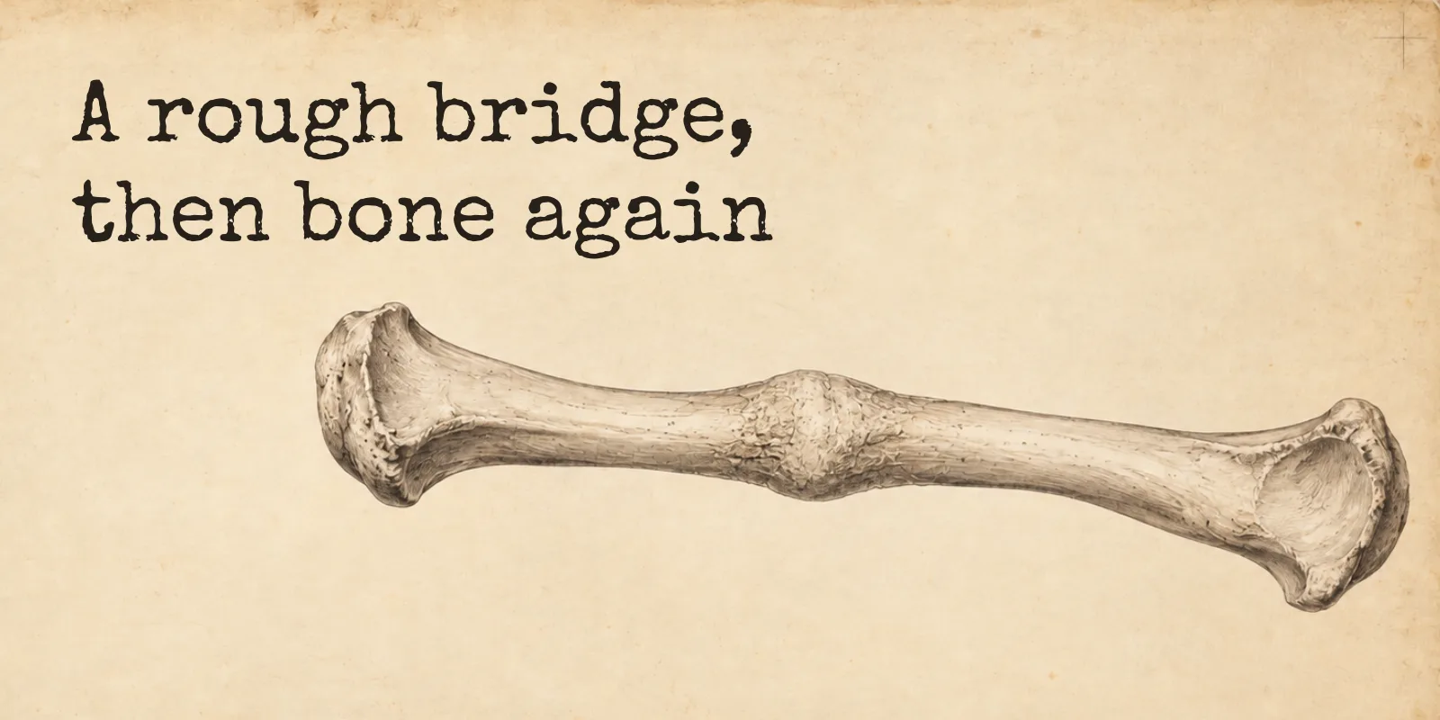
A break heals in four overlapping stages: a clot, a bridge of cartilage, a rough collar of new bone, then years of quiet reshaping. The part I did not expect is that bone is one of the few tissues in the body that repairs itself without leaving a scar.
A broken bone heals in four overlapping stages. Blood clots around the break within hours, a bridge of cartilage grows across the gap over the following weeks, that bridge is replaced by rough new bone over a few months, and then the body spends years quietly reshaping the repair until it looks like the bone that was there before.
The stages have names, and once you know them the phrases on an X-ray report stop being noise. A callus is the temporary bridge. Ongoing remodeling means the reshaping is still going, even though the cast came off a while ago.
The part that surprised me
Cut your skin and you get a scar, and the scar is not skin. I assumed a healed fracture worked the same way, that somewhere in the middle of the bone there was a patch of something second-rate holding the two halves together.
It does not work like that. A review of bone regeneration puts it about as plainly as a journal ever does: most fractures heal without forming scar tissue, and the bone is rebuilt with its original properties largely restored, so that the new bone eventually cannot be told apart from the uninjured bone beside it. Bone is one of the very few things in you that can do that.
The four stages are how it manages it.
Stage 1: the hematoma, in the first hours
A fracture tears the blood vessels running through the bone and the sleeve of tissue around it, so the first thing that happens is bleeding. About six to eight hours after the break, the blood clots into a fracture hematoma, and that clot is where everything else gets built.
Inflammatory cells arrive to clear away damaged tissue and dead bone. The acute inflammatory response peaks within the first twenty-four hours and is finished after about a week. New vessels start growing into the clot, and a soft, well-supplied tissue called granulation tissue forms in it within the first three to seven days.
Nothing here is holding the bone together yet.
Stage 2: the soft callus, a bridge of cartilage
The bridge starts going up sooner than you would think. Within about forty-eight hours of the break, cells inside the bone begin laying a fibrocartilage matrix between the two broken ends, and at the same time cells under the outer membrane build a second collar of cartilage and bone around the outside. That gives you an internal callus in the gap and an external callus wrapped around the break.
Together they are the soft callus, and it takes roughly two to four weeks to become a proper bridge. It is cartilage rather than bone, so it is flexible, and its job is to stop the ends moving enough for something stronger to be built inside it.
An X-ray taken in this window can still show the fracture line looking much as it did on day one, because cartilage that has not taken up any mineral barely registers on a plain film.
Stage 3: the hard callus, and why the new bone is rough
Now the cartilage gets replaced by bone, through the same process the skeleton used to build most of itself before you were born. It is called endochondral ossification, which sounds forbidding and just means bone forming where cartilage was.
The bone that appears first is woven bone, and it is put down in a hurry. Its collagen comes in irregular bundles rather than the neat arches of mature bone, and its cells are large, numerous and disorganized, which is what you get when the body prioritizes a bridge over a good bridge. Woven bone is also what a fetal skeleton is made of, for the same reason.
This is the hard callus. It usually takes two to four months for it to develop into a solid bony collar around the fracture, and it is the stage where an X-ray finally has something obvious to show: a cloudy, thickened area bridging the break. A report that says “abundant callus” is describing repair tissue doing its job, so it does not mean the bone has healed badly.
The collarbone is the honest example here, and it is a bone I have a soft spot for, because clavicle was one of the words I kept hearing on the forensics show that got me into all this. A broken clavicle heals close enough to the surface that you can feel the callus. The surgeons’ patient guidance says a large bump may develop over the fracture site as it heals, that it usually gets smaller over time, and that a small one often stays for good.
Stage 4: remodeling, which takes years
Once the two ends are solidly joined, the body starts taking its own scaffolding down.
Osteoclasts chew away the excess callus and the woven bone inside it, and osteoblasts lay down lamellar bone in its place, the mature kind with its collagen in orderly layers. The bulge shrinks. The internal architecture reorganizes itself along the lines the bone is actually loaded. Even the medullary cavity, the hollow channel down the middle of a long bone where the marrow sits, gets reopened through the repair.
Broad estimates put this at several months to years, and one review says flatly that remodeling may take years to finish. Taking the cast off is not the end of it. It means the repair has reached a point where a clinician is willing to change what you are allowed to do with it.
This is the same remodeling that goes on in an unbroken skeleton all your life, running at about 5 to 10 percent of your bones a year. A fracture just gives it somewhere specific to concentrate.
| Stage | What is happening | Roughly when |
|---|---|---|
| Hematoma and inflammation | Torn vessels bleed, the blood clots, damaged tissue is cleared, new vessels grow in | Clot at 6 to 8 hours, inflammation done by about 7 days |
| Soft callus | Cartilage bridges the gap inside and wraps the break outside | Starts within about 48 hours, takes 2 to 4 weeks |
| Hard callus | Cartilage is replaced by woven bone, giving a solid but rough bridge | Around 2 to 4 months |
| Remodeling | Woven bone becomes lamellar bone, the bulge shrinks, the marrow cavity reopens | Several months to years |
A word on those numbers, because they are softer than the rest of the article. The big references will not give you the middle of it. OpenStax puts a clock on the first two days and then stops. The two standard biology reviews describe the first week closely, hand you “may take years” for the end, and say nothing at all about the span in between.
The weeks and months above come instead from the engineering side of the literature, where you cannot build a model of a healing bone without a timeline, and where the same set of ranges gets passed from paper to paper in almost identical words. So take them as the shape of the thing rather than a schedule. If a page online gives you a firm number of weeks for a named bone, it knows something the reviews do not.
Why different books give different stages
You will find this described as three stages, four, or five. The events underneath are the same ones; what changes is where each book decides to draw its lines.
The reference The Bone Room checks itself against, OpenStax Anatomy and Physiology, walks through the whole thing without ever using the words soft and hard callus. A widely used clinical summary names its four stages hematoma formation, granulation tissue formation, callus formation and bone remodeling, and then says outright that there is considerable overlap between them. Split the first stage into bleeding and inflammation and you have five.
The overlap is the honest part. At two weeks there is granulation tissue, cartilage and the first mineral all present at once, in different parts of the same repair.
What an X-ray can and cannot tell you
Imaging runs a long way behind the biology. One recent review of fracture imaging reports that plain radiographs missed unmineralized callus in over 40 percent of cases that were healing perfectly well clinically, because a bridge with no mineral in it is close to invisible on a film. The same review notes that radiographs cannot show soft tissue changes, early healing, or how strong the bone actually is, and that two readers looking at the same early film often disagree.
So the sequence you can see and the sequence going on inside are not in step. A soft callus is real for weeks before it shows up. A report can still say “ongoing remodeling” long after the fracture is stable enough to use.
None of this will let you read your own X-ray report and work out how your own fracture is doing, and I am not qualified to help you try. It should at least make the words in it point at something.
Some fractures skip the callus entirely
Everything above describes secondary healing, which is what happens when a bone is held reasonably still but can still flex a little, as it can in a cast or a splint. It is by far the most common form, and the callus is its signature.
Hold the two ends together rigidly enough, with plates and screws, and the bone takes a different route. Under absolute stability the fragments knit directly across the gap, and there is little or no visible callus at all.
That is worth knowing before you compare X-rays with someone, because two fractures can both be healing correctly and look nothing like each other on film.
When the sequence does not run
Three words come up when it goes wrong, and they mean different things.
A malunion is a fracture where the pieces have grown back together in the wrong position. A nonunion is one where they have failed to join at all, given more than enough time; one clinical definition puts that at nine months from the injury with no sign of progress for the last three. Delayed union sits in between, healing that is taking longer than expected, and the sources are noticeably vaguer about where the line falls.
The things that push a fracture that way are mostly about supply and stability. Smoking and diabetes are named as the most important risk factors for nonunion, and a bone with a poor blood supply cannot heal, whatever else is done for it. Infection, inadequate fixation and too much movement at the break do the same.
Frequently asked questions
What are the four stages of bone healing?
Hematoma and inflammation, soft callus formation, hard callus formation, and remodeling. Some references split the first stage in two and describe five, and others use a different set of labels for the same events; all of them say the stages overlap.
How long does a broken bone take to heal?
There is no single answer, and the major references decline to give one. The broad ranges are inflammation over the first week, a soft callus at two to four weeks, a hard callus at around two to four months, and remodeling continuing for months to years. The bone involved, the fracture, and the person all change it, and only your clinician can say anything about a specific injury.
What is a callus on a broken bone?
The temporary bridge of repair tissue that grows across a fracture. It starts as cartilage, the soft callus, and is then replaced by woven bone, the hard callus, which the body later trims back and rebuilds as mature bone.
Does a broken bone grow back stronger?
Not permanently. The hard callus is bulkier than the original bone for a while, so the repair is thicker at that point, but remodeling then removes the excess and rebuilds the site to something close to the bone that was there before.
Can a broken bone heal in the wrong position?
Yes, and that is called a malunion: the ends join, but at an angle, rotated, or shorter than they were. It is distinct from a nonunion, where the ends do not join at all.
The thing I keep coming back to is that a cast holds still and the bone inside it does not. It throws up a rough bridge in a few months because a rough bridge is better than none, and then spends years taking that bridge down again and replacing it with something it is not embarrassed by.
Knowing which bone is doing it makes the whole business more interesting, which is roughly why I built the lab in the first place. It is free, and you can start with the big ones without knowing a single name.
Sources
Every claim above is checked against OpenStax Anatomy and Physiology 2e, StatPearls and other references on the NCBI Bookshelf, or a peer-reviewed review article. Two entries sit outside that group and carry no anatomical claim: a Cleveland Clinic page, for the plain-language definitions of malunion and nonunion, and the surgeons’ patient guidance from the AAOS, for the bump on a healing collarbone. The weakest citation here is the one carrying the week-and-month figures, which comes from the modeling literature rather than from a measurement; the article says so where it uses them. Where sources give different stage names, the text says so too.
Show all 13 sources
- OpenStax, Anatomy and Physiology 2e, 6.5 Fractures: Bone Repair: the clotting blood forms a fracture hematoma about six to eight hours after the fracture; within about 48 hours chondrocytes from the endosteum create an internal callus of fibrocartilaginous matrix while periosteal chondrocytes and osteoblasts create an external callus around the break; over the next several weeks osteoclasts resorb the dead bone and the cartilage is replaced by trabecular bone via endochondral ossification; the calli unite and compact bone replaces spongy bone at the outer margins.
- OpenStax, 6.4 Bone Formation and Development: in endochondral ossification bone develops by replacing hyaline cartilage; osteoblasts secrete osteoid, which calcifies; osteoclasts resorb the bone lining the medullary cavity.
- StatPearls, Fracture Healing Overview: the stages are hematoma formation, granulation tissue formation, callus formation and bone remodeling, with considerable overlap between them; granulation tissue forms within two weeks; primary healing occurs under absolute stability with strain below 2 percent, through Haversian remodeling; secondary healing occurs with non-rigid fixation at 2 to 10 percent strain, via endochondral healing.
- R. Marsell and T. A. Einhorn, The biology of fracture healing, Injury, 2011: a hematoma is generated immediately after the trauma; the acute inflammatory response peaks within the first 24 hours and is complete after 7 days; the second resorptive phase remodels the hard callus into a lamellar bone structure with a central medullary cavity; remodeling may take years to complete; direct healing requires anatomic restoration and rigid fixation, while indirect healing is the most common form.
- The stage timings, from the modeling literature: Computational modeling of human bone fracture healing affected by different conditions of initial healing stage, BMC Musculoskeletal Disorders, 2019, and A coupled computational framework for bone fracture healing and long-term remodelling, International Journal for Numerical Methods in Biomedical Engineering, 2022. Both give granulation tissue at 3 to 7 days, a cartilaginous soft callus in 2 to 4 weeks, a hard bony callus after 2 to 4 months, and restructuring over several months to years. Their wording is nearly identical, so treat them as one line of citation rather than two independent measurements; the article says as much.
- P. V. Giannoudis and colleagues, Bone regeneration: current concepts and future directions, BMC Medicine, 2011: unlike other tissues, the majority of fractures heal without the formation of scar tissue, and bone is regenerated with its pre-existing properties largely restored, the newly formed bone eventually indistinguishable from the adjacent uninjured bone.
- StatPearls, Histology, Bone: in woven bone the osteocytes are numerous, large and disorganized; in lamellar bone they are fewer, smaller, spindle-shaped and architecturally regular.
- Endotext, Anatomy and Ultrastructure of Bone: Histogenesis, Growth and Remodeling: woven bone is characterized by irregular bundles of collagen fibers and forms when bone is laid down very rapidly, during development and fracture healing; lamellar bone’s fibers form arches for optimal strength; woven bone is progressively replaced by mature lamellar bone during remodeling.
- Radiological Innovations for Monitoring Bone Regeneration and Fracture Healing, 2025: radiographs missed unmineralized callus in more than 40 percent of clinically healing cases; radiographs are limited in their ability to detect soft tissue changes, early-stage healing or biomechanical bone strength; interobserver variability affects interpretation, particularly in early healing when callus is nonmineralized.
- StatPearls, Bone Nonunion: a nonunion is a fracture that persists for a minimum of nine months without signs of healing for three months; the most important risk factors are smoking and diabetes; a bone with decreased blood supply cannot heal; infection and poor mechanical stability impair healing.
- Cleveland Clinic, Malunion and nonunion fractures: a malunion is where the fractured bone parts have grown back together but in the wrong position; a nonunion is where they have failed to grow back together despite having had more than enough time.
- AAOS OrthoInfo, Clavicle fracture (broken collarbone): a large bump over the fracture site may develop as the fracture heals, usually getting smaller over time, though a small bump often remains permanently.