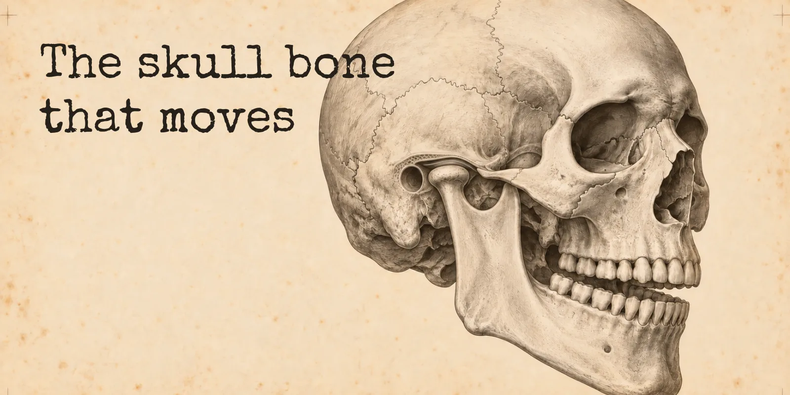
The mandible is your lower jaw, and it is the only bone of the skull that moves. It hangs from two joints just in front of your ears, and each of them hinges first and then slides, which you can feel for yourself with one finger.
The mandible is the lower jaw, and it is the only bone of the skull that moves. The other 21 skull bones are locked together into a single unit, and the mandible hangs beneath them from two joints, one just in front of each ear.
The name is a good one to start with, because it describes the job. It comes from the Late Latin mandibula, jaw, and before that from mandere, to chew.
There is one fair objection to “only bone that moves”, and it is the ear. The three tiny ossicles inside each middle ear pass sound along as vibration, so they move too. They just are not counted among the 22 bones of the skull, which the textbook lists separately as bones associated with the head, so the claim holds in the terms it was made. The post on how many bones are in the skull sorts out exactly who is in and who is out.
Where it sits, and what you can feel
The mandible is the largest bone of the face, and it counts as one of the 14 facial bones, so it is left out of the 8 cranial bones around the brain. It is U-shaped, and each side has two parts that meet at a corner.
The horizontal part is the body, which runs back from the chin and carries the lower teeth along its upper edge. The upright part is the ramus, from the Latin for branch, which climbs toward the ear. The corner where they meet is the angle of the mandible, the one you can find by running a finger back along your jawline until it turns upward.
At the top of each ramus the bone splits into two projections. The front one is the coronoid process, a flat blade that a chewing muscle pulls on. The back one is the condylar process, topped by an oval knob called the condyle, and that knob is the part that forms the joint.
You can find the condyle without knowing where to look. Put a fingertip just in front of the opening of your ear and open your mouth slowly, and you will feel something move forward under your finger. That is the condyle leaving its socket, and the clinical textbook on examining this joint notes that the movement is normally easy to feel.
One more thing I found surprising: the mandible starts out as two bones. At birth it is a left half and a right half, and they fuse into a single bone during the first year of life.
The joint in front of your ear
Each side of the jaw connects to the skull at a temporomandibular joint, the TMJ, named for the two bones it joins. The condyle of the mandible sits in the mandibular fossa, a deep oval hollow in the temporal bone just in front of the ear canal. In front of that hollow is a small downward bump called the articular tubercle.
Between the two bones there is a flexible disc that smooths the movement, and the whole joint is closed inside a capsule with fluid in it. That makes it a synovial joint, the same broad family as the knee and the shoulder.
I had always pictured the jaw as a hinge, and it is one, but only for the first part of the job. The Clinical Methods chapter on this joint says the condyle simply rotates in its hollow for roughly the first 25 millimeters of opening, measured between the front teeth. After that it glides forward along the slope of the tubercle, taking the disc with it, which is the forward movement you felt under your finger.
That glide is also why a jaw can dislocate. Open far enough, with a very big bite or a blow to the jaw, and the condyle can travel past the front of the tubercle and get stuck there.
One bone, two joints
Because the mandible is a single bone, the left and right joints can never act alone. Whatever one condyle does, the other has to accommodate, and StatPearls describes the two joints as coordinating so that the jaw moves in sync.
Pushing your jaw straight forward is the simple case. Both condyles slide forward together, pulled by the lateral pterygoid muscle on each side.
Moving the chin to one side is more interesting, because the two joints do different things at once. When only one lateral pterygoid pulls, the jaw swings toward the opposite side, so to move your chin to the left, the muscle on the right draws the right condyle forward while the left condyle stays more or less in its hollow and turns. The same logic shows up when something goes wrong: if one condyle cannot slide forward, the other still does, and the chin drifts toward the side that is stuck.
Why the other 21 stay still
Every other joint between the bones of the skull is a suture, which the textbook defines as an immobile joint between adjacent skull bones. At a suture the edges are joined directly by fibrous connective tissue, with no joint cavity between them, so there is no room for the kind of travel the jaw makes. OpenStax does allow that some sutures may permit slight movement between the cranial bones, but nothing like a jaw opening.
The words you will see on a report
A report that says mandible means the bone, from the chin back to both joints. These are the parts it may name.
| Term | Where it is | What is worth knowing |
|---|---|---|
| Body | The horizontal part, from the chin back to the angle | Its upper border, the alveolar process, holds the lower teeth |
| Symphysis | The midline of the chin | Where the left and right halves fused during the first year |
| Angle | The corner where the body turns up into the ramus | One of the most common places for the jaw to break |
| Ramus | The upright part on each side | The masseter, the main muscle used for chewing, attaches to its outer surface |
| Condyle, condylar process | The rounded knob at the top back of the ramus | The part that forms the joint with the temporal bone |
| Coronoid process | The flat projection at the top front of the ramus | Where the temporalis attaches; its front fibers help close the jaw and its back fibers pull the jaw backward |
| Mental foramen | A small hole on the outer face of the body, below the premolars | Where the nerve that gives feeling to the chin and lower lip comes out |
Mental here comes from the Latin for chin, so the mental foramen is the hole in the chin region and the mental protuberance is the chin itself.
That nerve has a longer route than the hole suggests. It enters the bone on the inner side of each ramus as the inferior alveolar nerve, runs forward through a canal inside the body sending branches to the lower teeth, and comes out at the mental foramen as the mental nerve. StatPearls places the foramen halfway down the body, most commonly in line with the gap between the first and second premolars.
When the jaw breaks
The mandible is one of the facial bones that breaks most often, and StatPearls names it alongside the nose as the most common facial fracture seen in emergency departments. The sites it gives as most common are the body, the condyle and the angle, while the ramus and the coronoid process are rarely involved.
It also tends to break in more than one place. StatPearls reports multiple fractures in more than half of cases and puts that down to the mandible’s ring-like structure, a U of bone held at both ends by the joints, so a blow at one point often shows up as a second break somewhere else.
These are the words that turn up in the notes on a broken jaw.
Malocclusion means the upper and lower teeth no longer meet the way they did, so the bite feels wrong. Because the lower teeth sit in the body of the mandible, a break that shifts part of the body shifts the teeth with it.
Trismus means the mouth will not open properly. The Clinical Methods chapter treats an opening of less than 35 millimeters between the front teeth as abnormal in an adult.
Sublingual hematoma is a collection of blood under the tongue, in the floor of the mouth. StatPearls says it suggests a mandible fracture that may not yet be obvious, and its article on body fractures goes further and calls bruising on the floor of the mouth a telltale sign of a fracture in the body.
Numbness of the lower lip and chin can happen when the break damages the inferior alveolar nerve on its way through the bone.
A break that tears the gum, the lining of the mouth or a tooth socket counts as an open fracture, because the bone is then exposed to the mouth, and StatPearls says antibiotics should be given to lower the risk of infection.
With a fracture just below the condyle, the jaw deviates toward the broken side, which is the same drift you would expect from one joint that can no longer slide.
How a particular fracture is treated is decided by the surgical team with the scans in front of them, and the location of each break matters a great deal to that decision.
Is the mandible the only movable bone in the skull?
Yes. OpenStax describes the mandible as the only moveable bone of the skull; the other 21 are joined at sutures, which are immobile joints. The tiny ear ossicles also move, but they are counted separately from the 22 skull bones.
What joint lets the mandible move?
The temporomandibular joint, one on each side, where the condyle of the mandible sits in a hollow of the temporal bone just in front of the ear. The joint rotates like a hinge for the first part of opening and then glides forward, and it also allows the jaw to push forward, pull back and move side to side.
Is the mandible a cranial bone or a facial bone?
It is a facial bone, the largest of the 14. The 8 cranial bones are the ones that surround the brain.
What are the signs of a broken jaw?
StatPearls lists trismus (difficulty opening the mouth), malocclusion (teeth that no longer meet properly), swelling and tenderness. Bleeding under the tongue and numbness of the lower lip or chin can also point to a fracture. A jaw injury needs examination and imaging to confirm.
If this has you running a finger along your own jawline, the lab is free. The mandible is one of the plates you meet in The Bone Room, a few specimens at a time, and you only get to claim it once you have named it on three separate days, the last time typed from memory.
Sources
Every anatomical claim above is checked against OpenStax Anatomy and Physiology 2e or StatPearls on the NCBI Bookshelf, the same references the lab uses, with the Clinical Methods textbook for the two measurements. Where two sources give different figures, the text says so.
Show all 13 sources
- OpenStax, Anatomy and Physiology 2e, 7.2 The Skull: the skull has 22 bones, 21 immobile and united into a single unit; the mandible forms the lower jaw and is the only moveable bone of the skull; 8 bones of the brain case and 14 facial bones; a suture is an immobile joint; the body, ramus (“branch”), angle, coronoid and condylar processes; the alveolar process anchors the lower teeth; the mandibular fossa lies just in front of the external acoustic meatus; the mental foramen as the exit for a sensory nerve to the chin; mental means chin; the two halves fusing in the first year.
- OpenStax, 9.6 Anatomy of Selected Synovial Joints: the TMJ joins the mandibular fossa and articular tubercle with the mandibular condyle; the articular disc smooths movement; opening combines hinge and gliding motion as the condyle and disc glide forward onto the tubercle; side-to-side and protraction/retraction; dislocation when the condyle moves in front of the tubercle.
- OpenStax, 9.2 Fibrous Joints: at a suture the bones are connected by fibrous connective tissue with no joint cavity; sutures are classed as synarthroses, though some may allow slight movement between the cranial bones.
- OpenStax, 11.3 Axial Muscles of the Head, Neck, and Back: the masseter as the main muscle used for chewing.
- OpenStax, 14.1 Sensory Perception: the ossicles pass vibration from the eardrum to the inner ear.
- OpenStax, 7.1 Divisions of the Skeletal System: the six ear ossicles and the hyoid listed as bones associated with the head, in addition to the 22 skull bones.
- StatPearls, Anatomy, Head and Neck, Mandible: U-shaped body and rami; mental foramen most commonly between the first and second premolars; mental nerve to the lower lip; inferior alveolar nerve entering the mandibular foramen and running in the mandibular canal with branches to the lower teeth; temporalis inserting on the coronoid process; two separate bones at birth, fused in the first year.
- StatPearls, Anatomy, Head and Neck, Temporomandibular Joint: capsule, synovial cavity and fluid, articular disc; rotation followed by translation during opening; coordination with the opposite joint; bilateral lateral pterygoid action protrudes the jaw and unilateral action deviates it to the opposite side.
- StatPearls, Anatomy, Head and Neck, Mastication Muscles: anterior and middle temporalis fibers elevate the mandible and posterior fibers retract it; the masseter elevates the mandible and inserts on the outer ramus.
- StatPearls, Mandible Fracture: nasal and mandible fractures as the most common facial fractures; body, condyle and angle as the most common sites, ramus and coronoid rarely involved; multiple fractures in more than 50% because of the ring-like structure; trismus, malocclusion, swelling and tenderness; sublingual hematoma suggesting an occult fracture; inferior alveolar nerve damage numbing the lower lip and chin; mucosal, gingival or tooth socket disruption making a fracture open, with antibiotics; a subcondylar fracture deviating the jaw toward the fractured side.
- StatPearls, Mandible Body Fracture: ecchymosis of the floor of the mouth as a pathognomonic sign of a body fracture; altered lower lip sensation in displaced body fractures.
- Walker, Hall and Hurst (eds), Clinical Methods, 3rd edition, The Temporomandibular Joint Examination: rotation for the first 25 mm of opening, then forward translation along the articular eminence; condylar movement is easily felt over the joint; opening under 35 mm abnormal in an adult; one condyle unable to translate makes the chin deviate toward that side.
- Online Etymology Dictionary, mandible: from Late Latin mandibula, jaw, from Latin mandere, to chew.